
Vitreous hemorrhage (VH) can be classified based on the location of the bleeding in relation to the retina and the vitreous. For medical advice or diagnosis, consult a professional. Here are five main types and their effects on vision:
- Preretinal Hemorrhage:
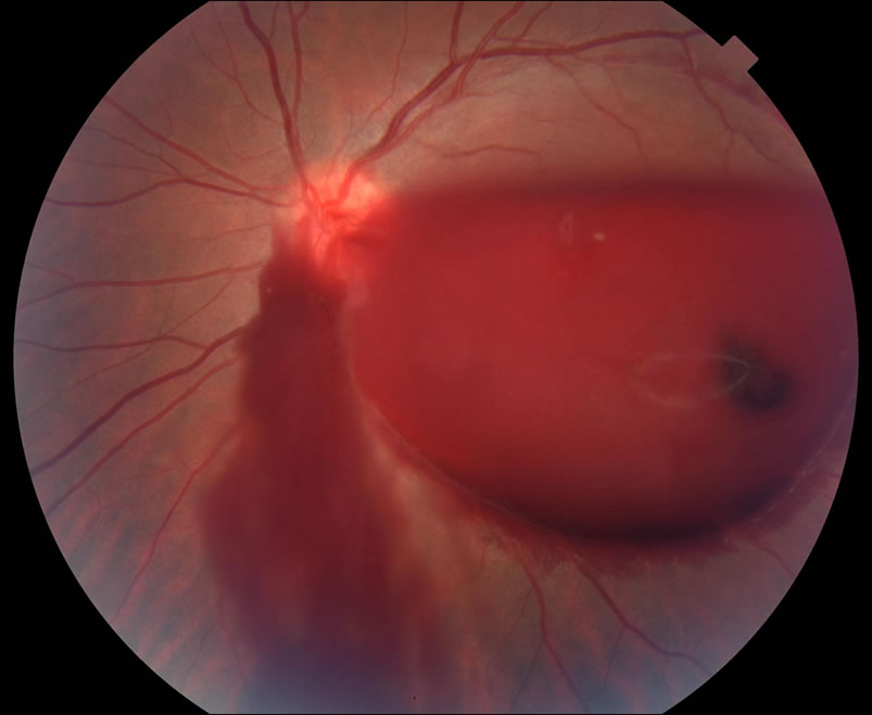
Location: Between the retina and the posterior hyaloid face (or under the internal limiting membrane).
Appearance: Boat-shaped or D-shaped hemorrhage due to blood settling with gravity.
Causes: Retinal vein occlusion, diabetic retinopathy, trauma, or Valsalva retinopathy (strain-induced bleeding).
Impact on Vision: Can cause significant vision loss if located over the macula, but may resolve over time. - Intravitreal Hemorrhage:
Location: Within the vitreous gel itself.
Appearance: Diffuse or clumped blood, causing a hazy red or dark view.
Causes: Retinal tear or detachment, proliferative diabetic retinopathy, trauma, or retinal vein occlusion.
Impact on Vision: Sudden vision loss, floaters, and hazy/cloudy vision; severe cases may obscure the entire visual field. - Sub-Internal Limiting Membrane (Sub-ILM) Hemorrhage:
Location: Between the internal limiting membrane (ILM) and the nerve fiber layer of the retina.
Appearance: Dark red or round hemorrhage that does not shift with gravity.
Causes: Terson’s syndrome (due to intracranial hemorrhage), trauma, or Valsalva retinopathy.
Impact on Vision: Can significantly reduce vision if over the macula; may require laser or surgery if persistent. - Subretinal Hemorrhage:
Location: Between the retina and the retinal pigment epithelium (RPE).
Appearance: Dark red, irregularly shaped hemorrhage beneath the retina.
Causes: Age-related macular degeneration (AMD), choroidal neovascularization, or retinal vascular diseases.
Impact on Vision: Can cause permanent central vision loss, especially if involving the macula. - Vitreous Base Hemorrhage:
Location: At the vitreous base (the area where the vitreous is most firmly attached to the retina).
Appearance: Blood localized at the peripheral retina, often detected during an eye exam.
Causes: Trauma, retinal tear, or vitreous detachment.
Impact on Vision: Usually does not affect central vision but may lead to floaters and peripheral vision disturbances.
Each type of hemorrhage has different implications for vision and may require different treatment approaches. A prompt ophthalmic evaluation is essential to determine the cause and management plan.
Signs and Symptoms:
1.Sudden vision loss (partial or complete)
2.Floaters (dark spots, cobwebs, red/black specks)
3.Hazy or cloudy vision (foggy, blurry sight)
4.Red tint or dark shadow in vision
5.Flashes of light (photopsia)
6.No pain or eye redness
7.Worsening vision in the morning (blood settles overnight)
Complications:
If left untreated, VH can lead to severe complications:
1.Persistent vision impairment (if bleeding does not clear)
2.Retinal detachment (if caused by a retinal tear)
3.Glaucoma (increased eye pressure due to blood blocking drainage)
4.Fibrosis and scarring (can cause retinal traction and detachment)
5.Recurrent hemorrhage (in conditions like diabetic retinopathy)
Prognosis:
Mild cases: Small hemorrhages may clear on their own within weeks to months.
Moderate cases: May take longer to resolve, requiring medical management.
Severe cases: May require surgery, especially if associated with retinal detachment or persistent bleeding.
Treatment:
- Conservative Management (Observation):
Small or mild VH cases may resolve on their own in weeks to months.
Bed rest with head elevation helps blood settle away from the central vision.
Regular monitoring with follow-ups and imaging (OCT, B-scan ultrasound if the retina isn’t visible). - Medical Treatment:
Anti-VEGF injections (e.g., bevacizumab, ranibizumab, aflibercept) if VH is due to proliferative diabetic retinopathy (PDR) or neovascularization.
Corticosteroids in cases of inflammatory causes.
Discontinuation of anticoagulants if deemed safe by the prescribing physician.
Control of underlying conditions like diabetes and hypertension. - Laser Therapy (Panretinal Photocoagulation – PRP):
Used when VH is due to diabetic retinopathy, retinal vein occlusion, or neovascularization.
Helps prevent recurrent bleeding by treating the underlying retinal ischemia. - Vitrectomy Surgery:
Removes the vitreous gel and hemorrhage, improving vision and preventing further complications.
Common in cases of diabetic retinopathy, retinal tears, or trauma.
This is for informational purposes only. For medical advice or diagnosis, consult a professional.
It’s important to have your eyes checked regularly by a qualified eye care professional.
Retinal (vitreous) hemorrhages requires careful evaluation and treatment by an eye care professional to prevent complications.
